Dedicated to delivering excellence in patient care by providing high quality clinical imaging services.
Committed to undertaking inspirational and impactful research, teaching the science and application of imaging and interventional radiology.
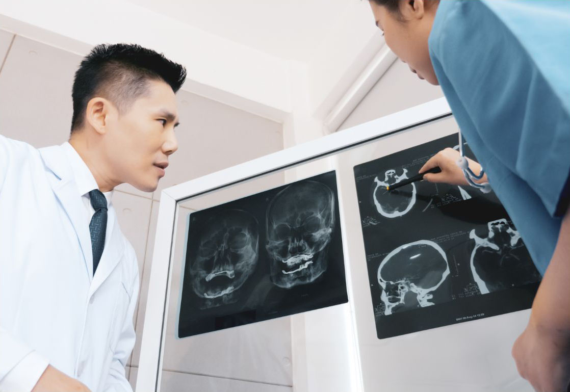
Established in 1986, we have long history of providing outstanding clinical service, research, and teaching in imaging and interventional radiology.
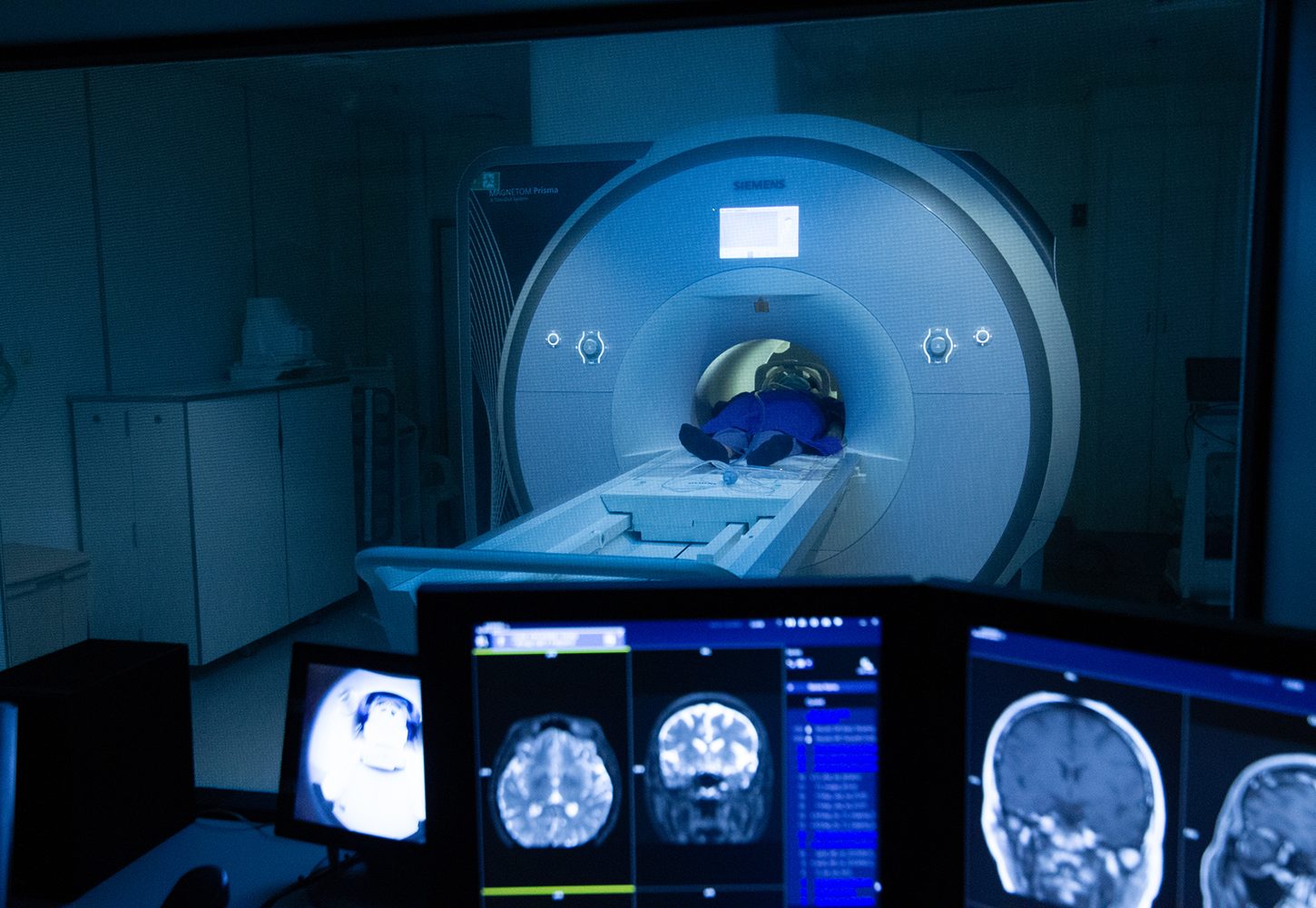
Equipped with state-of-the-art imaging equipment and technology, our Department provides comprehensive imaging services for diagnosis and treatment, as well as innovative technological development.
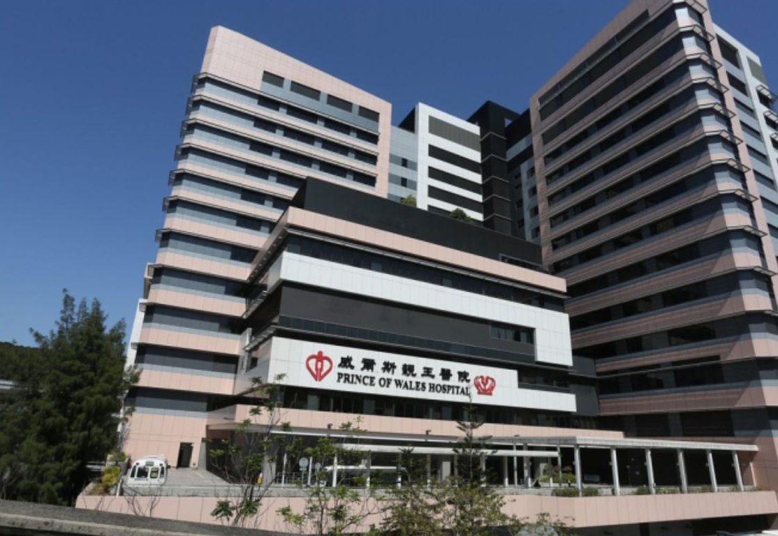
Located in the Prince of Wales Hospital and benefiting from collaboration with affiliated departments and hospitals, we provide a platform to promote knowledge transfer and groom future leaders.
We take great pride in being consistently ranked in the top 2 to 3 in Asia for the Best Global Universities in Radiology, Nuclear Medicine and Medical Imaging.
Highlights
MILESTONE
1986
Establishment of Department of Imaging and Interventional Radiology
2000
Installed first 1.5T MRI
2008
Installed first 3T MRI scanner
2012
Introduced Master of Science in Diagnostic Ultrasonography Program
2013
Introduced Diploma Program in Vascular & Interventional Radiology
2016
Established Research Centre for Medical Imaging Computing
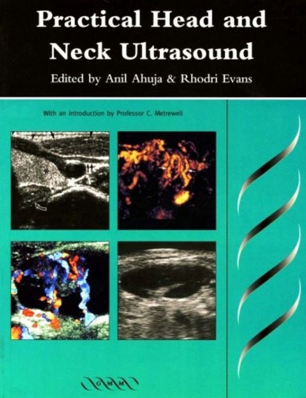
Ch. 1 Anatomy and Technique R. M. Evans
Ch. 2 Salivary Glands M. J. Bradley
Ch. 3 The Thyroid and Parathyroids A. T. Ahuja
Ch. 4 Lymph Nodes R. M. Evans
Ch. 5 Lumps and Bumps in the Head and Neck A. T. Ahuja
Ch. 6 The Larynx E. Loveday
Ch. 7 What the Surgeon Needs to Know, and Why D. W. Patton K. C. Silvester
Ch. 8 Fine-Needle Aspiration or Core Biopsy? N. J. A. Cozens L. Berman
Ch. 9 Carotid and Vertebral Ultrasonography S. Ho C. Metreweli
Imaging in Head & Neck Cancer
Contributor Anil Ahuja, Ann King
Publisher London : Greenwich Medical Media
Creation Date 2003
Language English
Identifier ISBN : 1841100900
Imaging in SARS
Contributor Ahuja, Anil T
Ooi, C. G. C. (Clara G. C.)
Publisher London ; San Francisco, Calif. : GMM
Creation Date 2004.
Language English
Identifier ISBN : 1841102199
Other title Imaging in severe acute respiratory syndrome
Case Studies in Medical Imaging: Radiology For Students and Trainees
Summary “This book is written as a system-based clinical-radiological review providing images from the latest available imaging modalities and it covers all major diseases that are envountered in everyday clinical practice. A problem-orientated approach is used. Each chapter contains a collection of clinical cases, each with a short clinical description and initial imaging followed by pertinent questions regarding the imaging findings (colour coded in red outline).” “This book is written by experienced radiologists currently working in undergraduate and postgraduate medical education. The cases are a collection of their illustrative teaching material.”–BOOK JACKET.
Publisher Cambridge ; New York : Cambridge University Press
Creation Date 2006
Language English
Identifier ISBN : 0521682940 (pbk.)
ISBN : 9780521682947 (pbk.)
Other title Medical imaging : radiology for students and trainees
Radiology for students and trainees
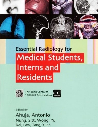
Essential Radiology for Medical Students, Interns and Residents
Abstract This is a comprehensive system-based review in clinical radiology, covering major diseases encountered in everyday clinical practice. A case-orientated approach is used, with high quality images from the latest available imaging modalities including MRI of the breast and cardiovascular system, CT colonography, amongst others. Each chapter is dedicated to a specific functional system and begins by outlining different clinical cases, each with a concise clinical history and initial imaging, followed by explanation of the pertinent imaging findings. The second part of each case contains a discussion on the list of important differential diagnoses to consider and the role of different imaging modalities in the presenting problem. The latter shows how information derived from different imaging modalities (including xrays, fluoroscopy, CT, MRI, nuclear medicine / PET-CT, angiograms, etc.) can help refine and prioritize imaging differentials. For each clinical case, the novel incorporation of cine images (accessible via QR codes) provides a new type of learning experience – one that simulates the reality of how medical imaging are interpreted using serial images with spatial reformatting rather than static planar images.
All Editor(s) List Anil T Ahuja, Gregory E Antonio, Jacqueline CM Sitt, Janice WL Yu, Simon SM Wong, Ryan CH Nung, Eunice YL Dai, Eric KC Law, Brian TY Yuen, Evelyn WK Tang