Introduction
Quantitative imaging (QI) is the extraction of quantifiable features from medical images for the assessment of normal or the severity, degree of change, or status of a disease, injury, or chronic condition relative to normal. Quantitative imaging includes the development, standardization, and optimization of anatomical, functional, and molecular imaging acquisition protocols, data analyses, display methods, and reporting structures. These features permit the validation of accurately and precisely obtained image-derived metrics with anatomically and physiologically relevant parameters, including treatment response and outcome, and the use of such metrics in research and patient care. Several practical aspects of QI include accuracy, precision, and clinical validity. The accuracy of a measurement describes how close the measurement is to a correct answer. Precision (repeatability and reproducibility) allows us to discriminate measurement error from biologic change. Finally, QI tools must ultimately have clinical validity; the results must be relevant to our practice, impacting patient care and improving outcomes. QI is increasingly applied in modern radiology practice, assisting in the clinical assessment of many patients and providing a source of biomarkers for a spectrum of diseases. QI is commonly used to inform patient diagnosis or prognosis, determine the choice of therapy, or monitor therapy response.
The aspects of Quantitative Imaging have been covered in other sections of this website, including: Artificial intelligence, Imaging biomarkers, Head and Neck, Neuroimaging, interventional radiology, Musculoskeletal Imaging, Body Imaging, Pediatric Imaging, Breast imaging. A few additional examples with the DDVD (diffusion derived vessel density) MRI biomarker are listed below:
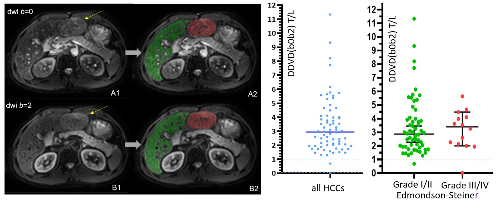
We applied DDVD to assess the perfusion of hepatocellular carcinoma (HCC). DDVD results (ratio of HCC DDVD to background liver DDVD equals around 3.0) approximately agreed with contrast agent dynamically enhanced CT/MRI literature data. We also demonstrated a slightly higher DDVD value for more malignment HCCs than for better differentiated HCCs.
reference:
Li XM, Yao DQ, Quan XY, Li M, Chen W, Wáng YXJ. Perfusion of hepatocellular carcinomas measured by diffusion-derived vessel density biomarker: Higher hepatocellular carcinoma perfusion than earlier intravoxel incoherent motion reports. NMR Biomed. 2024;37:e5125.

Correlation between DDVD and blood loss volume during surgery. A: pregnant women without PAS (placenta accreta spectrum disorders) and PAS patient data together, statistically significant correlation (p<0.0001) is noted between DDVD and blood loss volume during surgery. B: pregnant women without PAS and PAS patient data separately presented.
reference:
Lu T, Wang L, Li M, Wang Y, Chen M, Xiao BH, Wáng YXJ. Diffusion-derived vessel density (DDVD) computed from a simple diffusion MRI protocol as a biomarker of placental blood circulation in patients with placenta accreta spectrum disorders: A proof-of-concept study. Magn Reson Imaging. 2024;109:180-186.
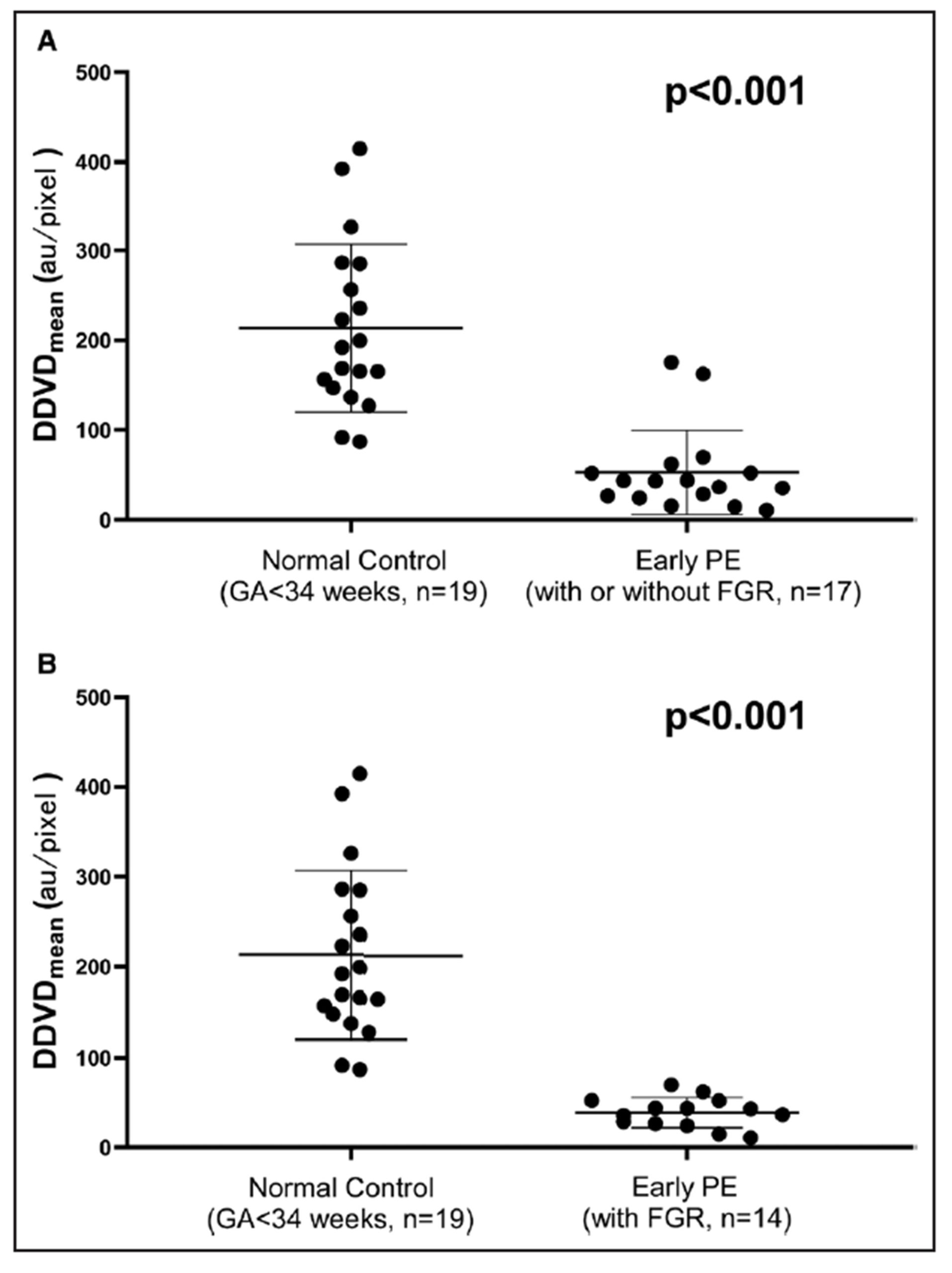
Box plots of DDVDmean in the control group and early preeclampsia (PE) group. A, Control group with gestational age (GA) <34 weeks and early PE group with or without fetal growth restriction. B, Control group with GA <34 weeks and early PE group with fetal growth restriction.
reference:
He J, Chen C, Xu L, Xiao B, Chen Z, Wen T, Wáng YXJ, Liu P. Diffusion-Derived Vessel Density Computed From a Simplified Intravoxel Incoherent Motion Imaging Protocol in Pregnancies Complicated by Early Preeclampsia: A Novel Biomarker of Placental Dysfunction. Hypertension. 2023;80:1658-1667.